Osteomalacia Explained: Causes, Symptoms, and Soft Bone Solutions
Osteomalacia is a metabolic bone disorder characterized by a fundamental problem: the softening and weakening of bones. Unlike conditions that primarily involve bone density loss, osteomalacia occurs when the bone matrix – the structural framework of the bone – fails to adequately mineralize. This critical process, vital for bone strength and rigidity, is most commonly disrupted by a significant deficiency in vitamin D, which plays a pivotal role in the body's absorption of calcium and phosphate.
Often confused with other bone conditions, it's essential to understand what sets osteomalacia apart. While it may share some superficial similarities with osteoporosis, the underlying issue is distinct. Osteoporosis involves a thinning of already mineralized bone tissue, making bones porous and fragile. In contrast, osteomalacia is a problem with the initial *hardening* of new bone. The body produces new bone tissue, but without proper mineralization, it remains soft, leading to increased bone fragility, pain, and a higher risk of fractures. To dive deeper into these distinctions, explore our comprehensive article on
Osteomalacia vs. Osteoporosis: Key Differences in Bone Health. This condition primarily affects adults, whereas a similar mineralization defect in children, impacting their growing bones and growth plates, is known as rickets.
Understanding the Causes of Osteomalacia: Why Bones Soften
The journey to softened bones is typically rooted in factors that disrupt the body's ability to process and utilize vital minerals. Identifying the cause is the first step toward effective treatment.
The Dominant Role of Vitamin D Deficiency
The most prevalent cause of osteomalacia is a severe and prolonged deficiency in vitamin D. This essential fat-soluble vitamin acts like a hormone, facilitating the absorption of calcium and phosphate from the gut into the bloodstream. Without sufficient vitamin D, even an adequate dietary intake of calcium and phosphate cannot be effectively utilized by the body to mineralize bone.
Several factors contribute to vitamin D deficiency:
- Insufficient Sunlight Exposure: Our skin produces vitamin D when exposed to ultraviolet B (UVB) rays from the sun. Limited time outdoors, living in regions with low sunlight intensity, use of strong sunscreens, or covering most of the skin can significantly impair vitamin D synthesis. Individuals with darker skin pigmentation also require more sun exposure to produce the same amount of vitamin D compared to those with lighter skin tones.
- Dietary Inadequacy: While some foods are fortified with vitamin D (milk, cereals) and fatty fish (salmon, mackerel) are natural sources, diet alone is often insufficient to meet vitamin D requirements without adequate sun exposure or supplementation.
- Malabsorption Disorders: Conditions that impair the absorption of fats in the digestive system can also hinder vitamin D absorption, as it's a fat-soluble vitamin. Examples include celiac disease, Crohn's disease, cystic fibrosis, and states following bariatric surgery.
- Liver and Kidney Disease: The liver and kidneys are crucial for converting vitamin D into its active form. Dysfunction in these organs can severely impair vitamin D metabolism.
To learn more about the critical link between this vitamin and bone health, refer to
Vitamin D Deficiency & Soft Bones: The Impact of Osteomalacia.
Other Significant Etiologies
While vitamin D deficiency is paramount, other factors can also lead to osteomalacia:
- Phosphate Deficiency: Phosphate is another key mineral for bone mineralization. Excessive loss of phosphate through the kidneys, often due to rare renal tubular disorders, can lead to phosphate deficiency and subsequent osteomalacia. Certain medications or prolonged use of aluminum-containing antacids can also interfere with phosphate absorption or increase its excretion.
- Hypocalcemia (Low Calcium): While often secondary to vitamin D deficiency, primary hypocalcemia can also contribute. This might stem from parathyroid gland dysfunction (e.g., hypoparathyroidism) which regulates calcium levels.
- Certain Medications: Some drugs can interfere with vitamin D metabolism or mineral balance. Anticonvulsants (like phenytoin) are known culprits, as are certain chemotherapeutic agents.
- Oncogenic Osteomalacia: This rare form is triggered by specific tumors (often benign) that secrete fibroblast growth factor 23 (FGF23). FGF23 inhibits phosphate reabsorption in the kidneys and vitamin D activation, leading to severe phosphate wasting and osteomalacia.
Risk factors for osteomalacia are broad and include elderly age, darker skin pigmentation, obesity, prolonged indoor lifestyles, and geographical locations with limited year-round sunlight. These factors collectively highlight the global prevalence, which is often underestimated.
Recognizing the Signs: Osteomalacia Symptoms
The symptoms of osteomalacia tend to develop gradually and can often be subtle or mistaken for other musculoskeletal conditions, delaying diagnosis. Understanding these signs is crucial for early intervention.
Chronic Bone Pain
This is one of the most common and debilitating symptoms. The pain is typically:
- Diffuse and Aching: Often described as a dull, persistent ache rather than sharp pain.
- Location: Commonly affects weight-bearing bones such as the lower back, pelvis, hips, thighs, and ribs. Pain can also be felt in the arms and feet.
- Worsens with Activity: The discomfort usually intensifies with movement, physical activity, or pressure on the affected bones. It may improve slightly with rest.
- Tenderness: Bones may be tender to the touch, making even gentle pressure uncomfortable.
Muscle Weakness and Mobility Issues
Proximal muscle weakness, particularly in the legs and shoulders, is another hallmark symptom. This weakness can manifest as:
- Difficulty Rising: Struggling to get up from a seated or squatting position.
- Waddling Gait: A characteristic walking pattern caused by weak hip muscles.
- Frequent Falls: Increased instability and muscle weakness contribute to a higher risk of falling.
- Difficulty Climbing Stairs: A common complaint due to weakened leg muscles.
Fractures and Pseudofractures
Due to the softened nature of the bones, individuals with osteomalacia are highly susceptible to fractures, often with minimal or no trauma. These are termed
pathological fractures. A specific type of radiological finding, known as
Looser's zones or pseudofractures, are linear areas of demineralization that appear on X-rays, often perpendicular to the bone cortex, particularly in the femur, pelvis, and ribs. These are essentially incomplete fractures where the bone has failed to properly heal.
Other Associated Symptoms
In severe cases, or when hypocalcemia is pronounced, patients may experience:
- Muscle Cramps or Spasms: Due to low calcium levels.
- Numbness or Tingling (Tetany): Particularly around the mouth or in the extremities, indicating severely low calcium.
- Fatigue: A general feeling of tiredness due to the overall metabolic imbalance and chronic pain.
The cumulative impact of these symptoms can significantly impair a person's quality of life and independence.
Diagnosing Osteomalacia: The Path to Clarity
A comprehensive approach involving clinical evaluation, laboratory tests, and imaging is crucial for an accurate diagnosis of osteomalacia.
Clinical Evaluation
A thorough medical history helps identify risk factors, medication use, and symptom progression. A physical examination may reveal bone tenderness, muscle weakness, and a waddling gait.
Laboratory Tests
Blood tests are essential for confirming osteomalacia and distinguishing it from other bone disorders:
- 25-Hydroxyvitamin D: The most important marker. Levels typically fall below 20 ng/mL, often below 10 ng/mL in severe deficiency.
- Calcium: Often low (hypocalcemia) or borderline low.
- Phosphate: Frequently low (hypophosphatemia).
- Alkaline Phosphatase (ALP): Usually elevated. This enzyme is produced by bone cells and its increased levels reflect the body's attempt to repair and remodel poorly mineralized bone.
- Parathyroid Hormone (PTH): Often elevated as the body tries to compensate for low calcium by stimulating PTH release, which in turn tries to raise calcium by resorbing bone and increasing kidney reabsorption.
Imaging Studies
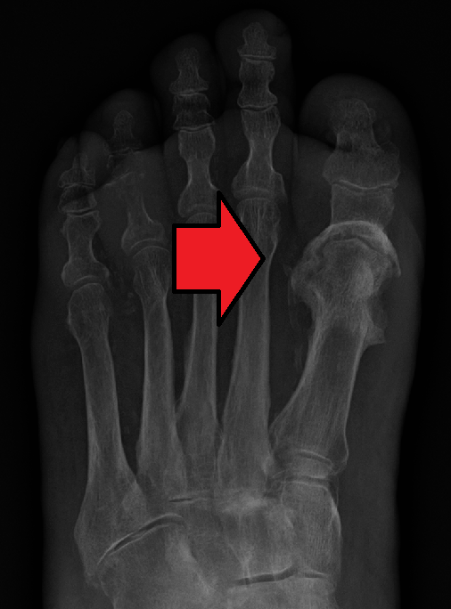
- X-rays: Can reveal characteristic findings like Looser's zones (pseudofractures). Generalized bone rarefaction (reduced density) may also be present.
- Dual-Energy X-ray Absorptiometry (DEXA) Scan: While not specific for osteomalacia, DEXA scans may show reduced bone mineral density (BMD), which is also seen in osteoporosis. It's useful for assessing overall bone health but needs to be interpreted in context with other tests.
Bone Biopsy: The Gold Standard
In ambiguous or complex cases, a bone biopsy, typically taken from the iliac crest (hip bone), remains the gold standard. Under a microscope, the biopsy will show characteristic widened osteoid seams – layers of unmineralized bone matrix that are abnormally thick, confirming the diagnosis of osteomalacia.
Effective Solutions: Treatment and Management
The good news is that osteomalacia is largely treatable, especially when the underlying cause is identified and addressed. The primary goal of treatment is to correct the mineral deficiency and promote proper bone remineralization.
High-Dose Vitamin D Supplementation
For cases stemming from vitamin D deficiency, treatment typically begins with high-dose vitamin D supplementation (cholecalciferol or ergocalciferol). The initial dose can be quite high, followed by a maintenance dose once levels normalize. This must be done under medical supervision, with regular monitoring of blood levels of vitamin D, calcium, and phosphate to ensure efficacy and prevent toxicity.
Calcium and Phosphate Supplementation
Alongside vitamin D, calcium and phosphate supplements may be prescribed if dietary intake is insufficient or if deficiencies are significant. It's crucial that these minerals are provided in conjunction with vitamin D to allow for proper absorption and utilization.
Addressing Underlying Conditions
- Malabsorption: If malabsorption is the cause, treating the underlying gastrointestinal disorder (e.g., managing celiac disease with a gluten-free diet, controlling Crohn's disease with medication) is paramount.
- Renal Disorders: For renal tubular disorders causing phosphate wasting, specific phosphate supplements and sometimes active vitamin D metabolites may be used under nephrologist guidance.
- Medication Review: If certain medications are contributing to the problem, alternatives may be considered, or managing their side effects.
- Oncogenic Osteomalacia: Treatment involves identifying and surgically removing the tumor. If removal is not possible, medications that block FGF23 action may be used.
Pain Management and Physical Therapy
While the underlying cause is being treated, pain relief is important. Non-steroidal anti-inflammatory drugs (NSAIDs) may be used for bone pain. Physical therapy can help strengthen muscles, improve mobility, and reduce the risk of falls, contributing to a better quality of life during recovery.
Prevention and Proactive Bone Health
Prevention of osteomalacia often boils down to ensuring adequate vitamin D, calcium, and phosphate levels throughout life.
- Sensible Sunlight Exposure: Aim for 10-30 minutes of direct sun exposure (unprotected skin on arms and legs) a few times a week, depending on skin type, location, and time of year. Be mindful of sun safety to prevent skin damage.
- Dietary Choices: Incorporate vitamin D-fortified foods (milk, yogurt, cereals) and natural sources like fatty fish (salmon, tuna, mackerel) into your diet. Ensure a calcium-rich diet with dairy products, fortified plant milks, leafy greens, and certain nuts.
- Supplementation: If dietary intake or sun exposure is insufficient, discuss vitamin D and possibly calcium supplementation with your doctor, especially if you have risk factors for deficiency.
- Regular Health Check-ups: Individuals with malabsorption disorders, kidney disease, or those on certain medications should have their vitamin D and mineral levels monitored regularly.
- Awareness of Symptoms: Don't dismiss persistent bone pain or muscle weakness. Early detection and intervention can prevent significant complications.
Conclusion
Osteomalacia is a serious but often treatable condition that results from the inadequate mineralization of bone, leading to softened, weakened bones. While it can be debilitating, causing chronic pain, muscle weakness, and an increased risk of fractures, an accurate diagnosis through blood tests and imaging, sometimes confirmed by bone biopsy, paves the way for effective treatment. The cornerstone of therapy typically involves correcting underlying vitamin D, calcium, or phosphate deficiencies, often through supplementation and addressing any contributing medical conditions. By understanding its causes, recognizing its symptoms, and pursuing appropriate medical care, individuals can restore their bone health, alleviate pain, and significantly improve their quality of life. If you suspect you or a loved one might be experiencing symptoms of osteomalacia, consult a healthcare professional without delay.